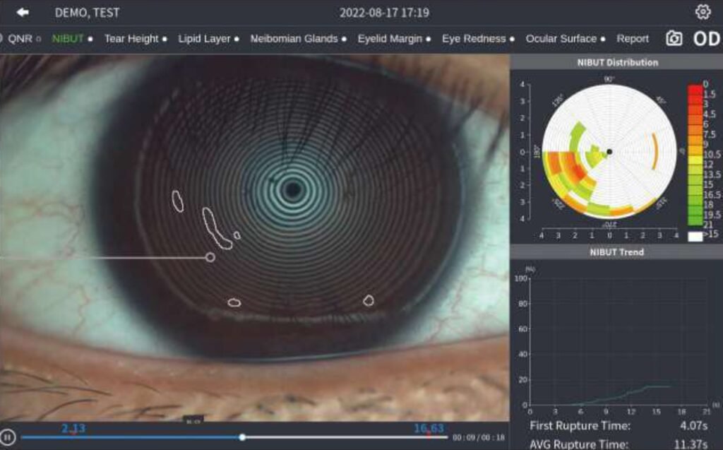
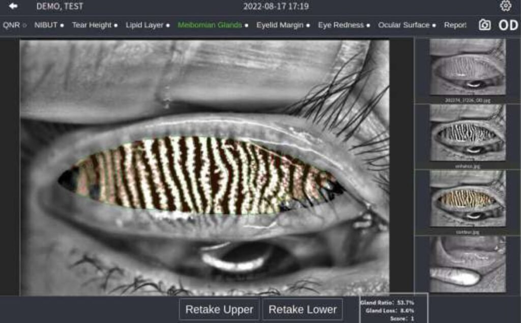
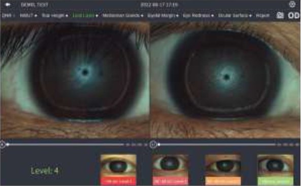
Lipid Layer Thicknes
Observe dynamic lipid layer and distribution by video recording compared with standard templates. It's helpful for judging MGD.
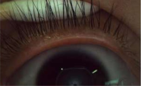
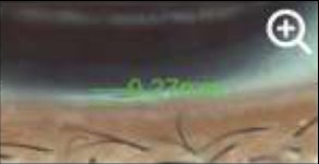
Eyelid Margin
The high resolution image supports zoom in to meet examination requirements of overall shape of eyelid margin and its slight change.
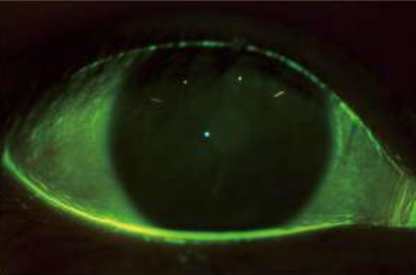
Cornea Sodium Fluorescein Staining
Specially designed built-in yellow filter, working with cobalt-blue illumination improves image contrast of cornea sodium fluorescein. Effectively increases posi- tive rate of early corneal epithelial staining.
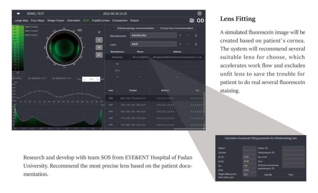
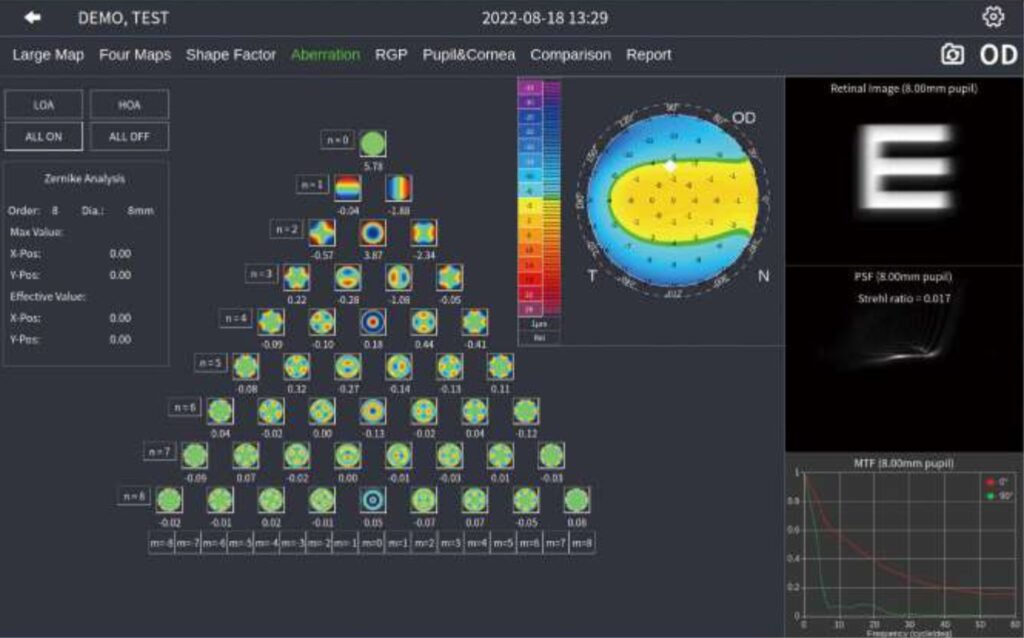
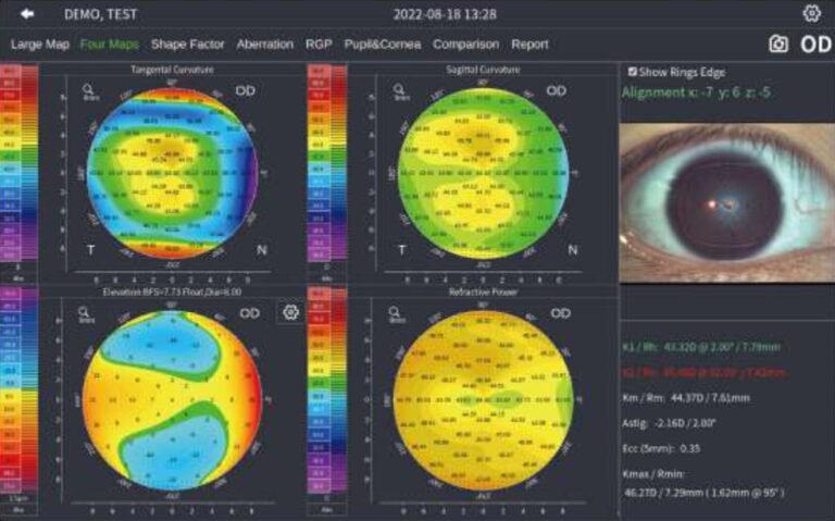
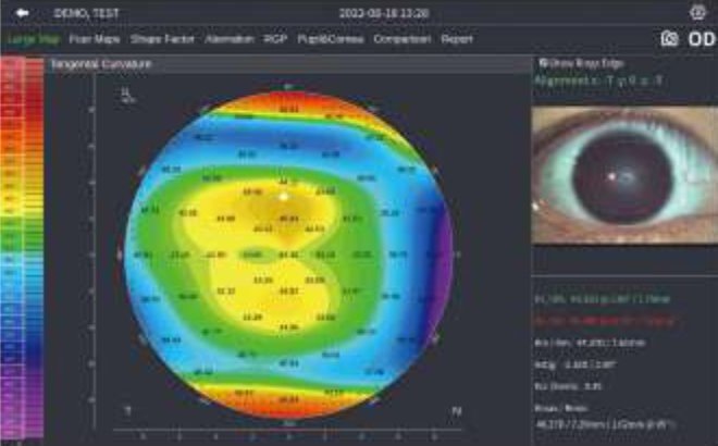
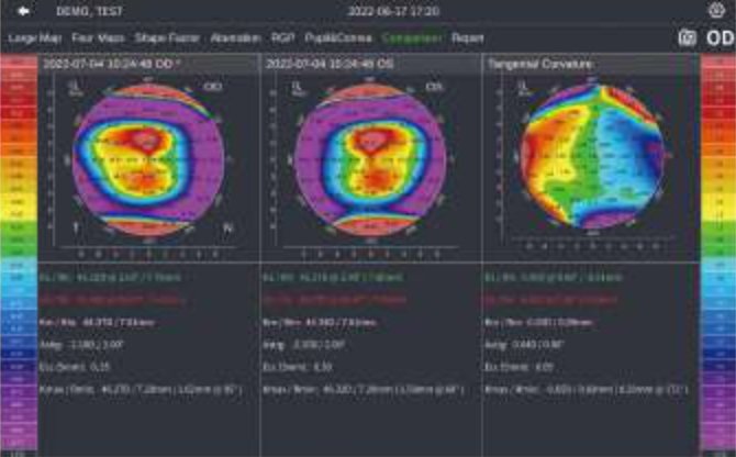
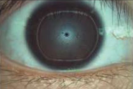
Topography
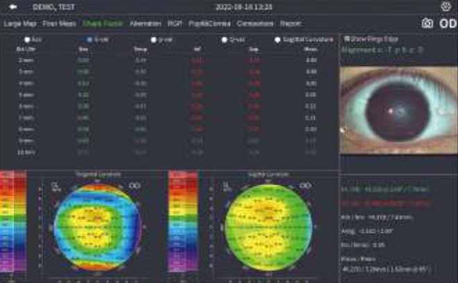
Shape Factor
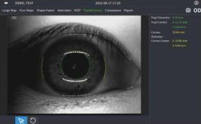
Pupil & Corneal Diameter Measurement