The ESL 5HI Dry Eye Diagnostic System is an advanced tool designed to diagnose dry eye conditions. It uses high-resolution imaging to assess tear film quality and other ocular surface characteristics, enabling eye care professionals to identify the severity of dry eye and tailor treatment accordingly. The system offers accurate, non-invasive testing for better patient care.

Platform for Comprehensive Ocular Surface Examination

Dry eye diagnosis/Anterior Segment Photography/Lens fitting/Patient management/Telemedicine

Guided examination: providing a comprehensive report covering 7 dry eye diagnosis.
Non-invasive examination, Quantitative data.

Full-automatic Firefly digital module, easy operation without parameter settings.

High quality optics and built-in yellow filter efficiently increase the accuracy of lens fitting.


Professional 1/1.8-inch sensor and 2.4µm pixel, real-time playing and storage.

Smart patient management system, DICOM supported.

Dry eye diagnostic system

Easy Pathogenic Diagnosis provides guidance for customized treatment.

Automatic Classification of Meibomian Glands

Unique built-in infrared lighting system provides a larger scope capture of Meibomian Glands, adjustable depth of field and aperture enables more vivid images. Precise diagnosis of Dry Eye caused by MGD is guaranteed with the help of automatic Meibomian Glands loss classification.

Increase positive rate of early corneal epithelial staining

Built-in yellow filter along with cobalt-blue filter increases the contrast of Sodium Fluorescein Staining image.

HD Optical System

Resolution is up to 200 lp/mm, providing more details of the pathologies.

Full Cornea Dry Eye Analysis

By Placido ring projection system with visible light, the examination scope is up to 8mm cornea diameter. Examination of the tear film outside of pupil center has the same significance for the diagnosis of Dry Eye.

Fully automatic Firefly digital module

Firefly Digital module is specially designed for anterior segment examination, no parameter settings required (automatic exposure, auto white balance, auto focus), with adjustable depth of field and wide dynamic range. 5 Mega Pixels video output allows high examination efficiency.

Simple Design + Simple Operation

The smart design largely saves space for clinicians compared to other bulky camera systems. We have preset many camera parameters so the user does not need to adjust settings before using the device. The user can operate the machine immediately once the installation has been finished. The device has the following automatic functions for photo shooting and processing when equipped with our software:

  1. Wide Dynamic Range
  2. Meibomian Glands Examination
  3. Auto Exposure
  4. Auto Gain
  5. Auto White Balance
  6. Auto DS/OD Indicator

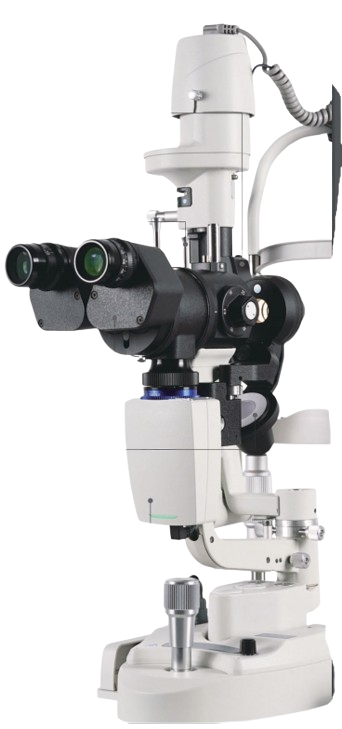
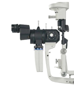
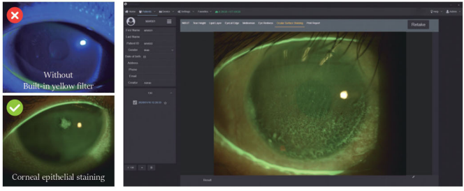
High sensitivity: The drill is still clear and sharp under weak light.

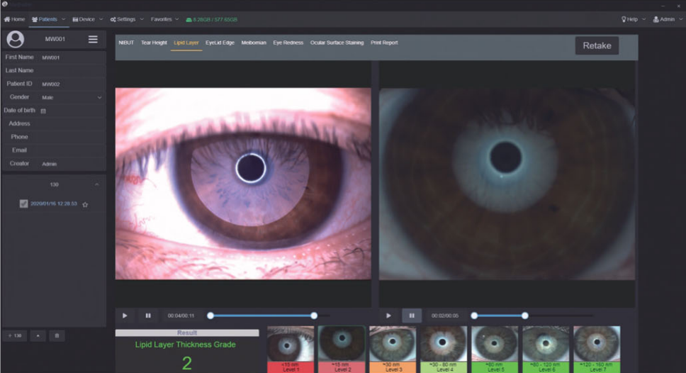
Wide dynamic range: This added feature improves the visibility, provides flexibility, presented with more realistic and evenly distributed color.

Software Features

Patient Management System

Get patient record perfection enables clinics to build a convenient Patient Management system. The patient management system records symptoms and key indicators enabling an easy method to build and manage data all the time. The software supports DICOM which makes the images captured by Fluvius be easily integrated into the hospital’s medical system.

Functional Image

Pathology Analysis Clinicians can measure the retina characteristics with our powerful software tools and change the contrast and brightness of the images. Clinicians also analyze the complex retinal images at all times to assist you at speedways pathology.

Orthokeratology Lens Fitting Assistance

The optometrists can capture and record high resolution fluorescein images of lens fitting and real-time video without a recording time limit. By comparing the different lens fitting effects, the optometrist can show and educate patients which lens is most suitable for them.

Customized Auto Exposure Value Setting

Clinicians can customize auto exposure values according to the image demand and save as templates for future capturing purposes. Also, the printing report can be customized according to clinician’s needs.
Due to various causes of Dry Eye Disease, traditional examination is difficult to find out the cause and quantify for the diagnosis. Eyevis Dry Eye Diagnostic System can provide standardized examination and quantified causes evaluation for Dry Eye Disease.

Dry eye classification from the 2007 DEWS Report

Functions

AI Non-Invasive Break Up Time

After taking one video, it brings out automatic results of NIBUT and Tear Meniscus Height.

Grade 0 Normal: First Rupture Time: 10 s, Average Rupture Time: 14 s

Grade 1 Warning: First Rupture Time: 6-9 s, Average Rupture Time: 7-13 s

Grade 2 Dry eye: First Rupture Time: ≤5 s, Average Rupture Time: ≤7 s

All identities the break-up area and analyzes NIBUT automatically. Fully automatic analysis system provides efficient quantified evaluation for the overall stability of tear film. It automatically acquires the first break-up time, average break-up time, break-up distribution/break-up area percentage curve, and time distribution.
Eyevis adopts Placido ring projection system with visible light to do NIBUT examination; the examination scope is up to 8mm cornea diameter which brings much more comprehensive diagnosis outcome.

The non-invasive examination avoids the irritation brought by the traditional Cornea Sodium Fluorescein Staining.

AI Non-Invasive Tear Meniscus Height

AI identification system depicts Tear Meniscus area and measures the tear height automatically.

Evaluate tear secretion amount and continuity objectively.

More efficient and less irritation compared with the traditional Schirmer’s test.

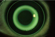
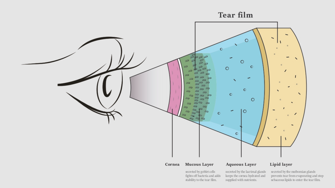
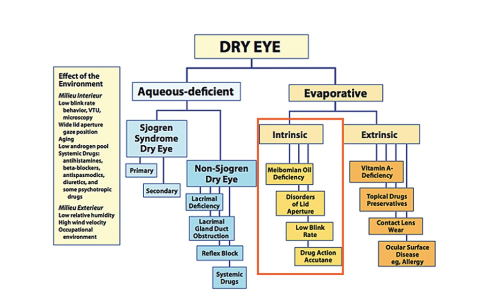
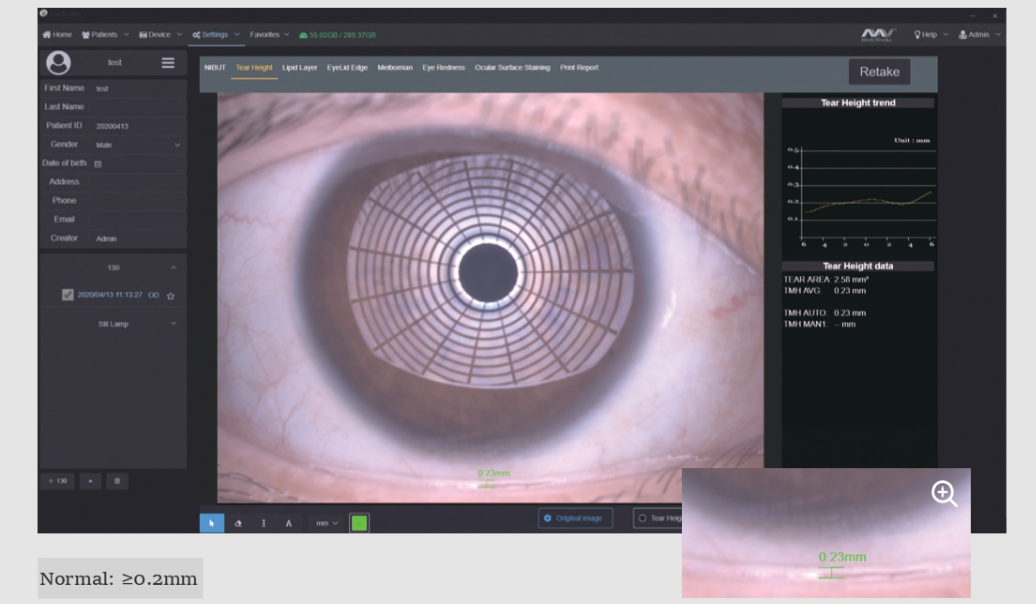
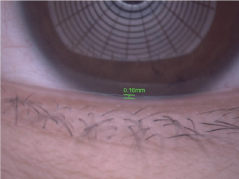
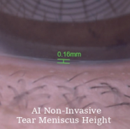
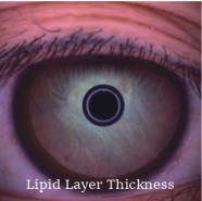
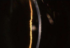
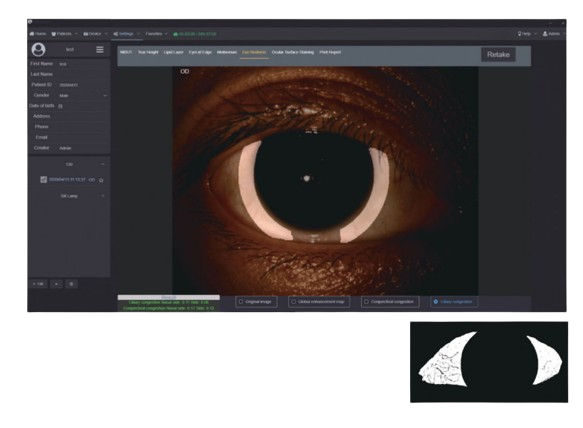
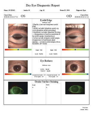
Insufficient tear secretion

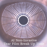
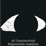
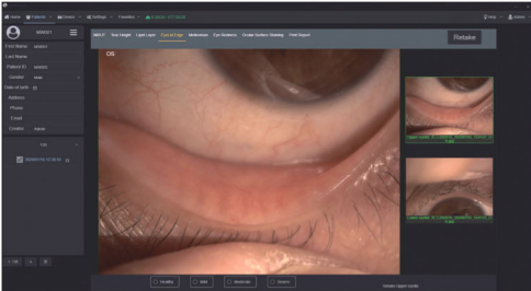
Abnormal dynamics and conjunctival chalasis

Evaluation of Meibomian Glands Function.

Automatic classification system provides precise and quantified diagnosis of DES caused by meibomian glands dysfunction. With built-in infrared lighting system, doctors can observe larger image scope of the Meibomian Glands. Adjustable depth of field makes the glands more prominent and distinguishable against the background.

Grade 0: No Meibomian Glands Loss
Grade 1: Meibomian Glands Loss < 1/3
Grade 2: Meibomian Glands Loss 1/3-2/3
Grade 3: Meibomian Glands Loss > 2/3

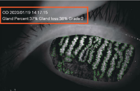
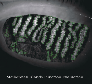
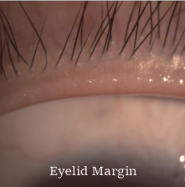
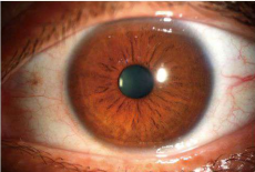
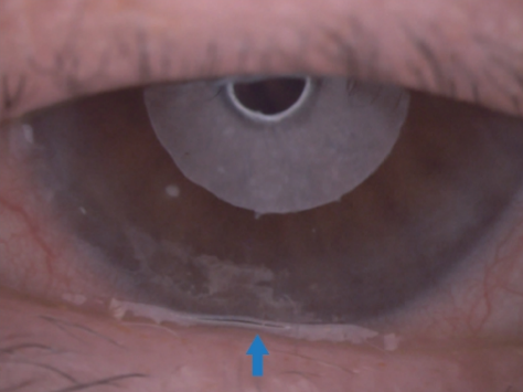
Meibomian glands loss

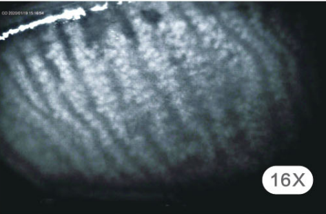
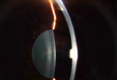
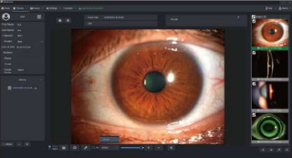
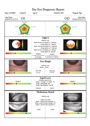
Image of Meibomian Glands under high magnification.

Lipid Layer Thickness

White ring projection system ensures a larger examination area compared to Placido ring. By comparing with the standard grading template and recording the Lipid Layer thickness, it is helpful for judging MGD.

Grade 1: < 15 (Unit: nm)
Grade 2: ~ 15
Grade 3: = 30
Grade 4: = 30-80
Grade 5: = 80
Grade 6: = 80-120
Grade 7: = 120-160″**

Eyelid Margin

1. Normal including (Ophthalmic embolism bright, transparent)
2. Mild including (gland cap crown – glandular prominent)
3. Moderate including (glandular fat plug – disappearance of the marginal mucosa, hyperkeratosis)
4. Severe including (uneven margins, disappearance of the meibomian glands – posterior margin Blunt round, thickening, new blood)

Eyevis professional design of optical system is capable of providing HD digital image that remains clear and sharp even zoom in, meets the examination requirements of the overall shape of eyelid margin and its slight change.

AI Analysis of Conjunctival Hyperemia

The unique AI identification system can identify and calculate percentages of conjunctival congestion and ciliary congestion and evaluate severity of eye congestion.

Cornea Sodium Fluorescein Staining

Effectively increases positive rate of early corneal epithelial staining. Built-in yellow filter along with cobalt-blue filter makes the corneal sodium fluorescein images more clearly.

Dry Eye Comprehensive Evaluation Report

Smart Patient Management system

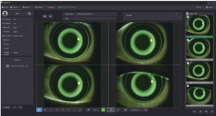
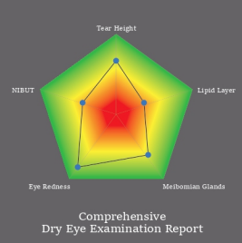
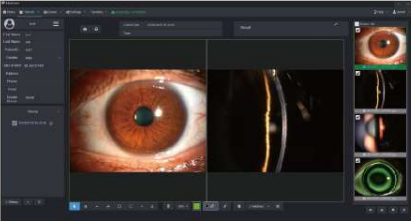
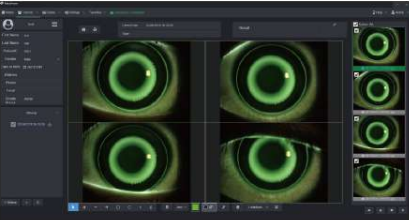
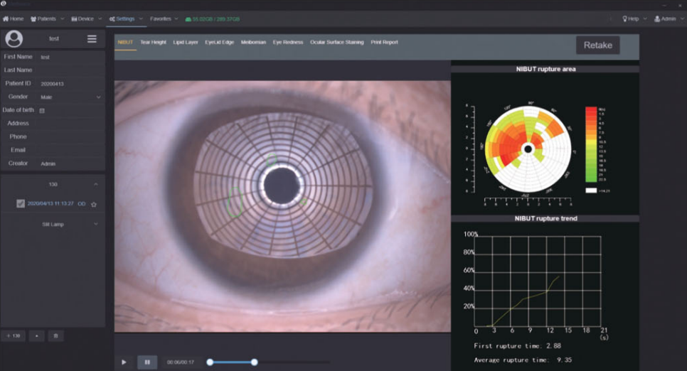
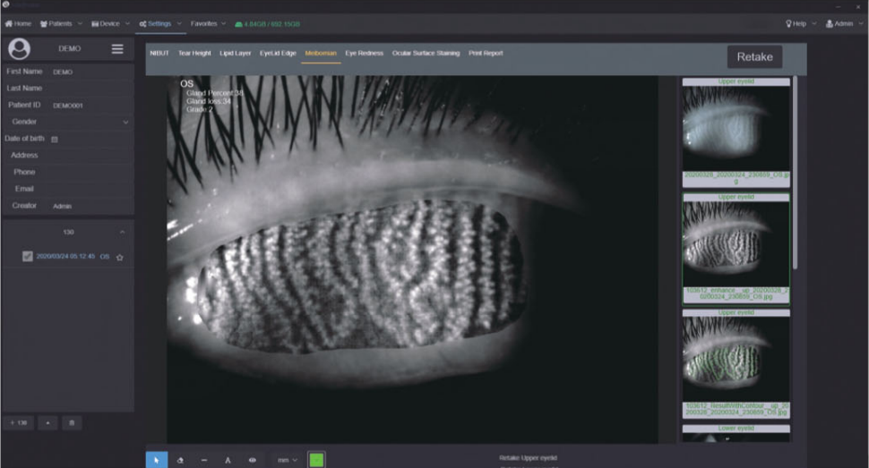
Comparison of Patient records

Supports repeated comparison among medical records to evaluate treatment and guide customized treatment plan.

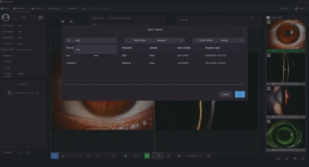
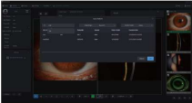
Patient Management system allows doctors to build and edit medical records. Quickly search the patient case by keywords. Doctors can note patients’ situation via the software. This DICOM-supported system enables Mediview to connect with medical systems in hospitals."**

Specification

Specifications ESL5H ESL3H
Microscope Type Galilean Type
Magnification Change Revolving drum 5 steps Revolving drum 3 steps
Total Magnification 6.3X, 10X, 16X, 25X, 40X 10X, 16X, 25X
Optical Resolution 2700-N lp/mm (200 lp/mm)
Eyepieces 12.5X
Angle between Eyepieces 52mm-80mm
Pupillary Adjustment 52mm-80mm
Diopter Adjustment -8D to +8D
Field of View Ø38.2mm, Ø22.3mm, Ø14mm Ø22.3mm, Ø14.3mm, Ø8.9mm
Slit Illumination Slit Weight 0-14mm continuous (slit becomes a circle at 14mm)
Slit Length 1-14mm continuous
Aperture Diameters Ø14mm, Ø10mm, Ø5mm, Ø3mm, Ø2mm, Ø1mm, Ø0.2mm
Slit Angle 0°-180°
Slit Inclination 5°, 10°, 15°, 20°
Filters Heat-absorbing filter, ND filter, Red-free filter, Cobalt blue filter, Built-in yellow filter
Lamp LED
Luminance 2150 kx
Power Supply Input Voltage 100V-240V
Input Frequency 50Hz/60Hz
Rated Current 1.2A
Output Voltage LED 3V, Fixation 15V
Packaging Dimension 740mm x 450mm x 530mm (L/W/H)
Gross Weight 23kg
Net Weight 17kg

Reach Out