Artificial Intelligence in Optical Coherence Tomography (AI-OCT) enhances retinal imaging by using machine learning to analyze OCT scans quickly and accurately. AI automates the detection of eye diseases like glaucoma, diabetic retinopathy, and macular degeneration, improving diagnosis, treatment monitoring, and early detection. This technology enables faster, more precise analysis, reducing human error and helping clinicians make informed decisions.
Product leading Al screening technology Empowering eye CT detection
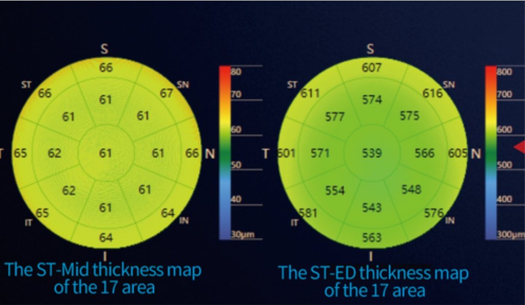
2) Automatic stratification measurement for choroid membrane
Accurate detection for myopia development intervention
3) Comprehensive functions, to meet a variety of needs
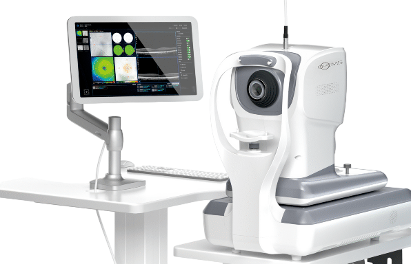
OCT functions such as fundus imaging, anterior segment imaging, SLO fundus imaging, eye tracking, and imaging of cloudy liquids are equipped
4) Al eye disease screening system, to locate the abnormal images
Automatically identify common fundus lesions and conduct risk assessment
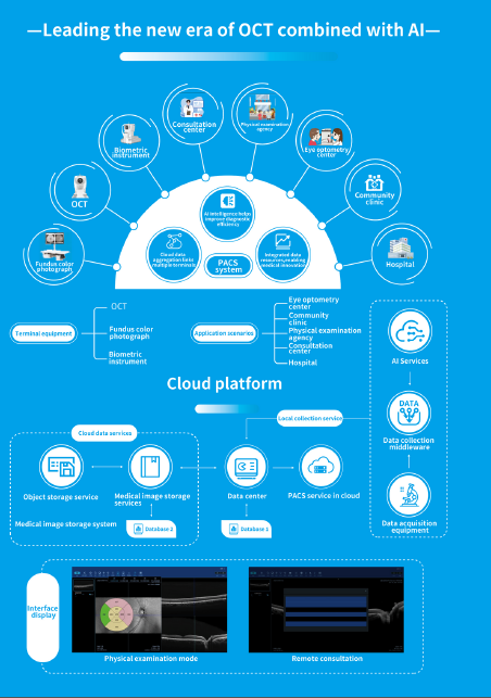
5) Cloud data archiving for information linkage
Cloud data aggregation with multiple terminals, efficient information linkage, instant transmission and reading, and high-speed information exchange
Al diagnosis, one-click enabling optometry detection / eye disease screening
Accuracy
0%
Eyevis OCT provides artificial intelligence disease screening systems.The systems can facilitate rapid fundus screening at the basic optometry center and improve diagnostic efficiency and accuracy. The systems can issue the diagnosis report with one button, with the accuracy of up to 97%. The systems are suitable for primary healthcare and optometry industries. At the same time, the Al Diagnosis Cloud Platform is built to realize data linkage
Data Source: From over 10,000 People real scene test
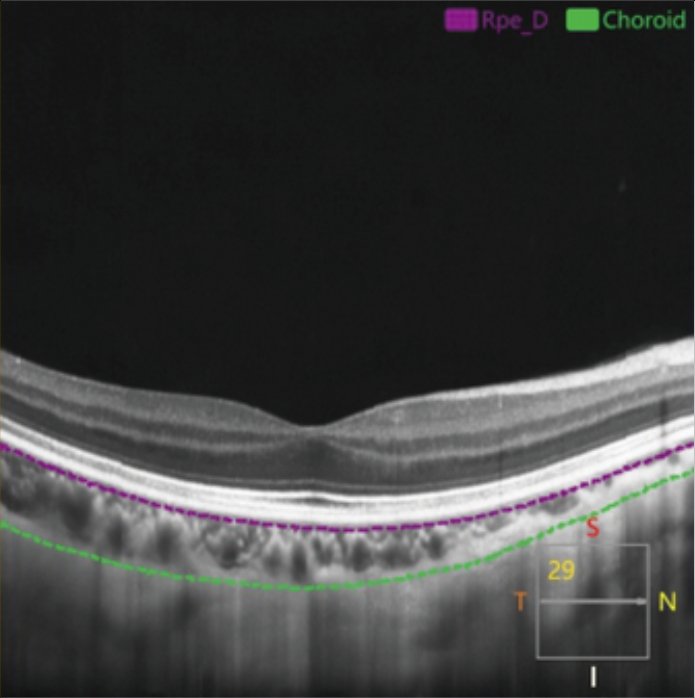
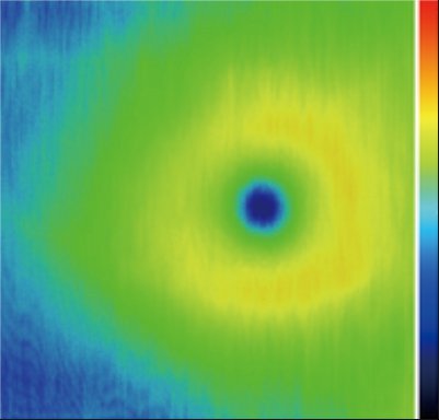
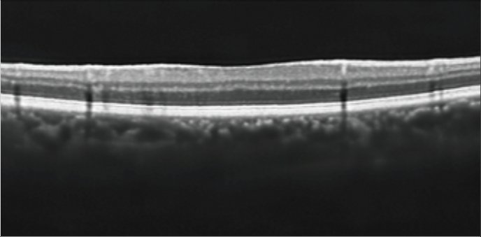
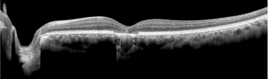
Automatic thickness measurement, no manual layering required
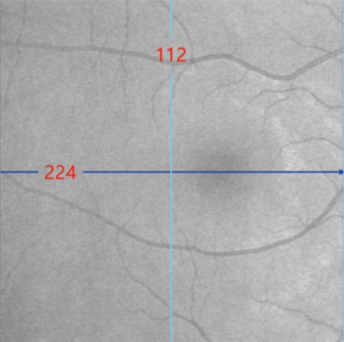
OCT functions include multiple scanning modes such as line scanning, grid scanning, and three-dimensional scanning, Macular fovea choroidal thickness (SFCT), choroidal subarachnoid thickness map and mean choroidal thickness in the macular region can be measured.
Thickness monitoring
before and after myopia prevention and control
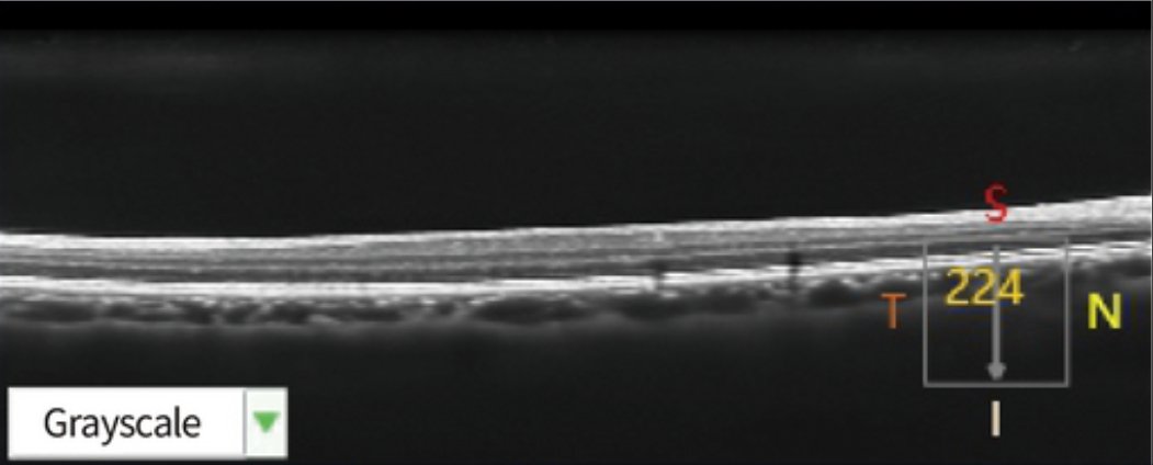
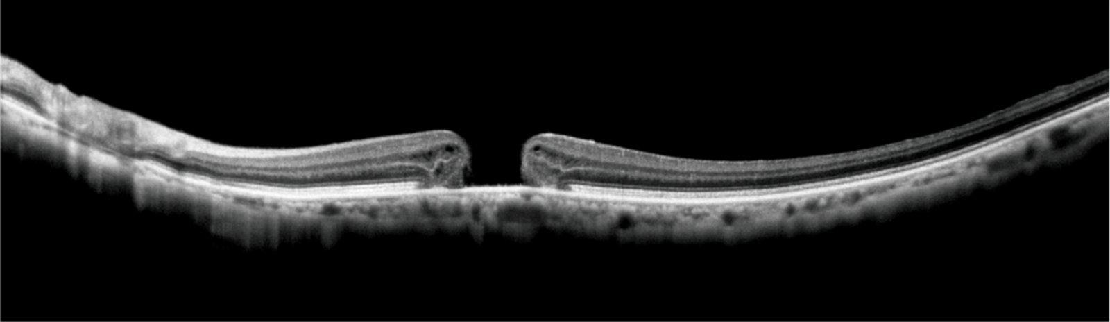
Choroidal thickness is directly related to myopia. The higher the diopter, the thinner the choroidal thickness. Measuring choroidal thickness through OCT can effectively evaluate the therapeutic effect of myopia prevention and control measures. Such as evaluating the effect of corneal reshaping lenses, functional lenses, low concentration atropine, and feed light meter on changing choroidal thickness.
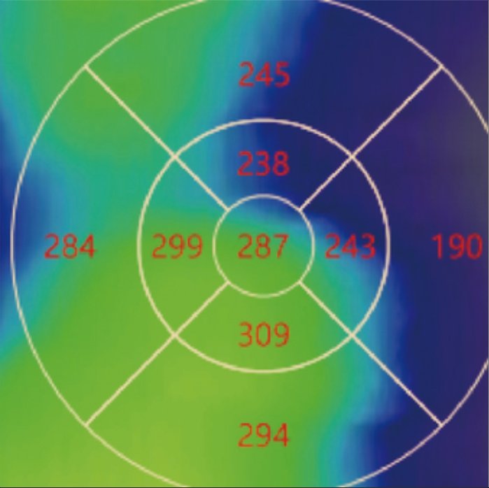
Choroidal thickness(µm)
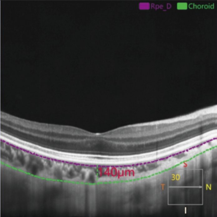
Macular fovea choroidal thickness:140µm
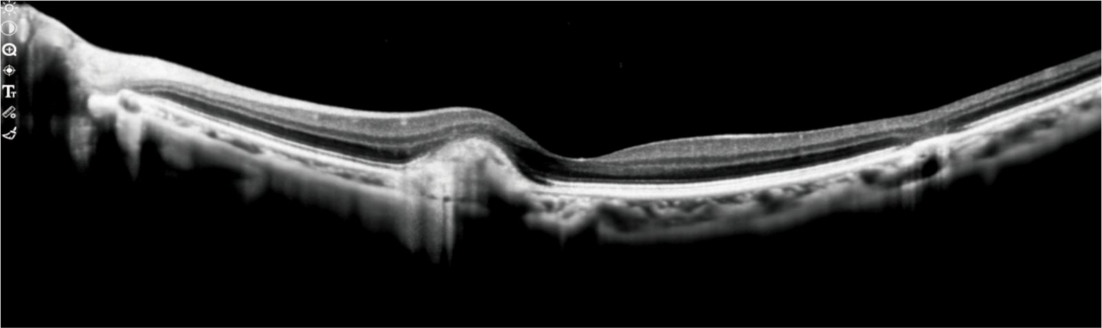
Report of high myopia choroid
Autofocus, less inspection time and easier operation
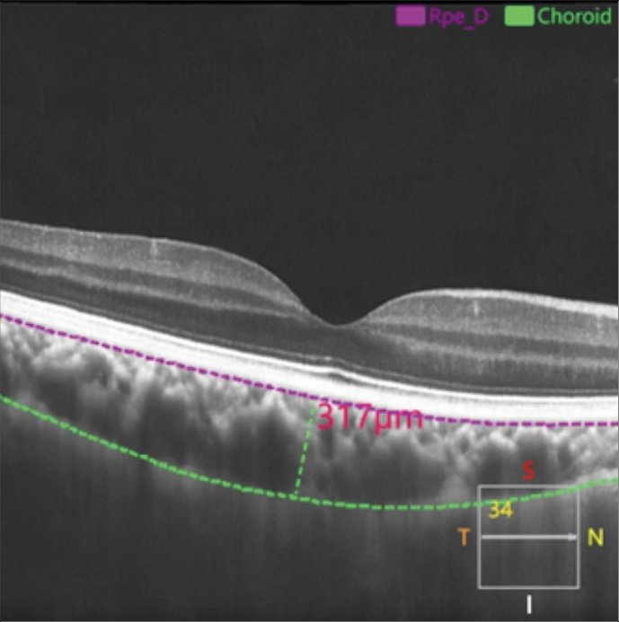
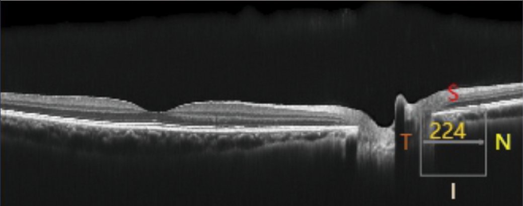
Al layering, automatic calculation of choroidal thickness
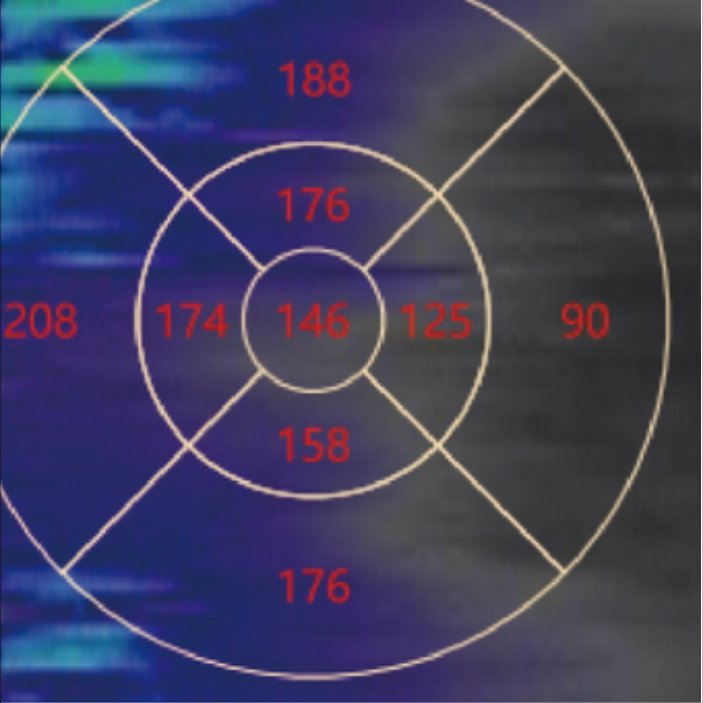
Automatic presentation of choroidal thickness and distribution map
Choroidal thickness(µm)
Macular fovea choroidal thickness:317µm
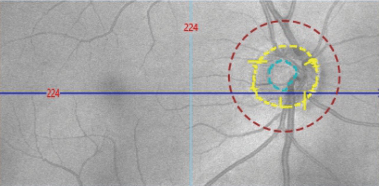
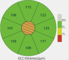
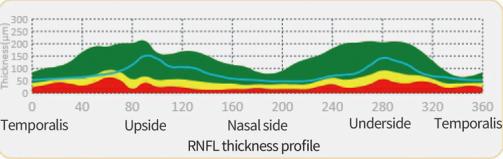
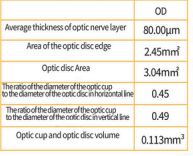
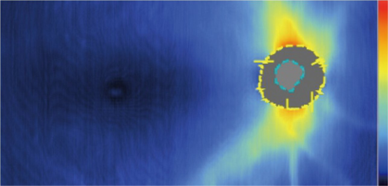
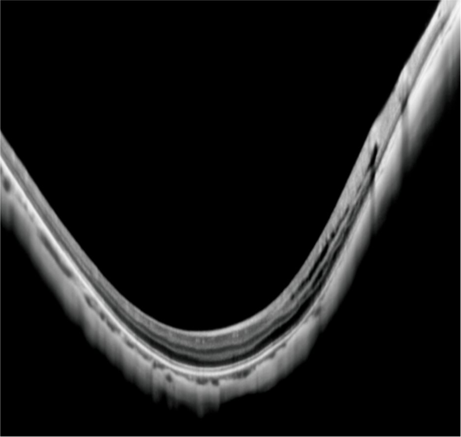
Glaucoma detection pattern
Glaucoma analysis -optic disk RNFL
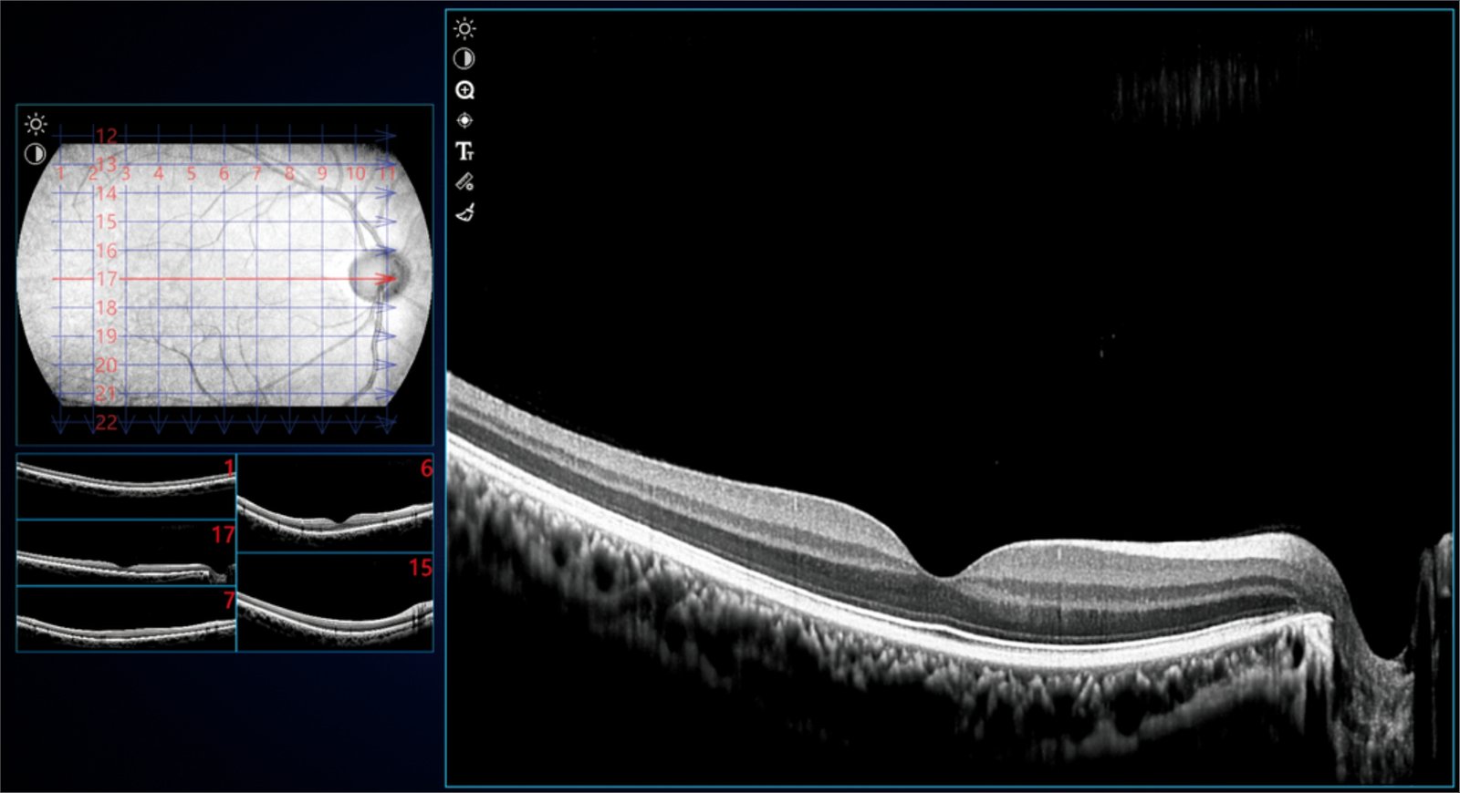
Single acquisition for 12mmx6mm Large range of fundus volume images
Thickness analysis and quantitative analysis can be performed on the macular area and optic disc area
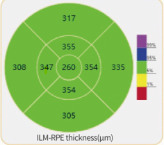
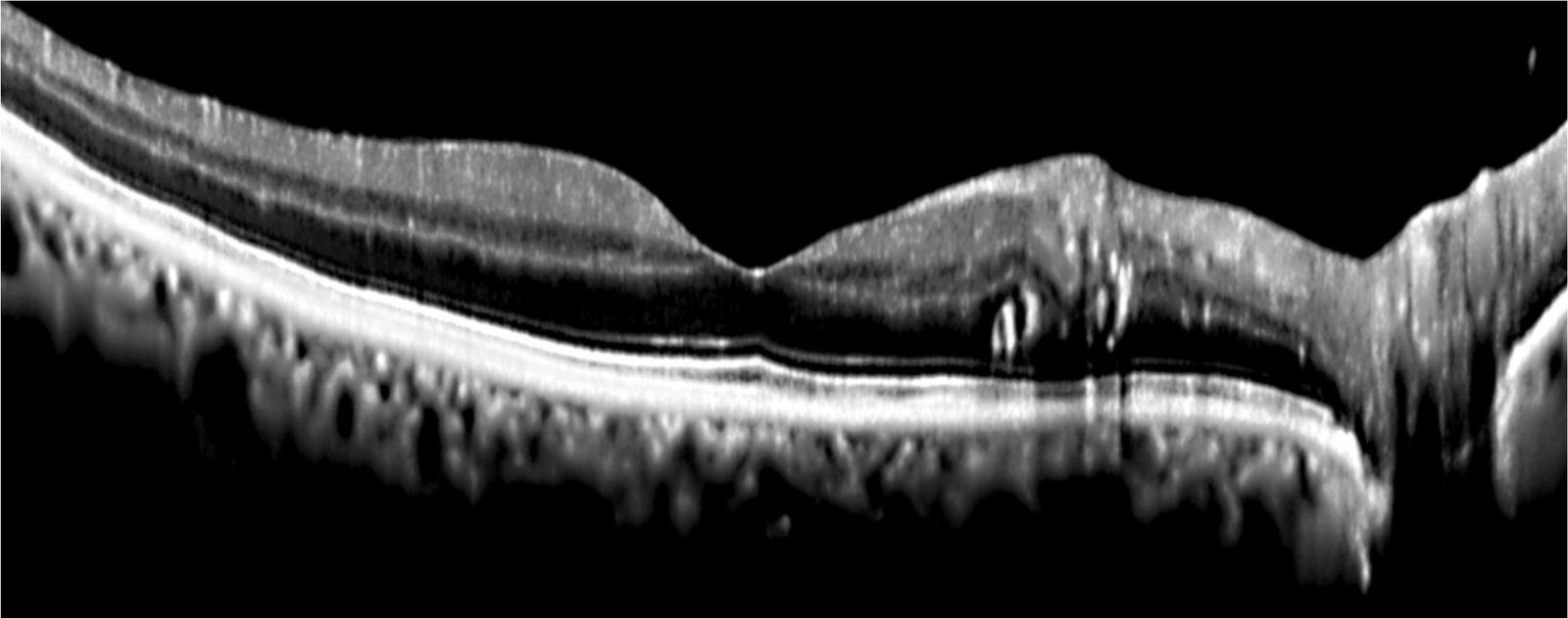
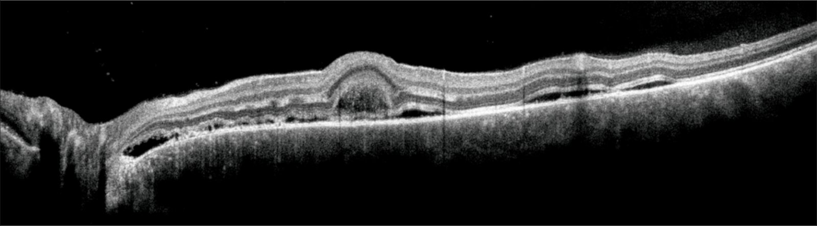
Macular detection mode
3D visualization, automatic thickness analysis
Clear stratification, without any loss of detail
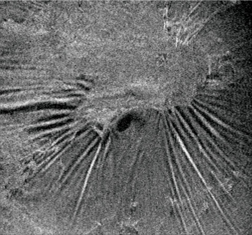
Multi-line scanning mode
more comprehensive scope
3D scanning analysis of macula
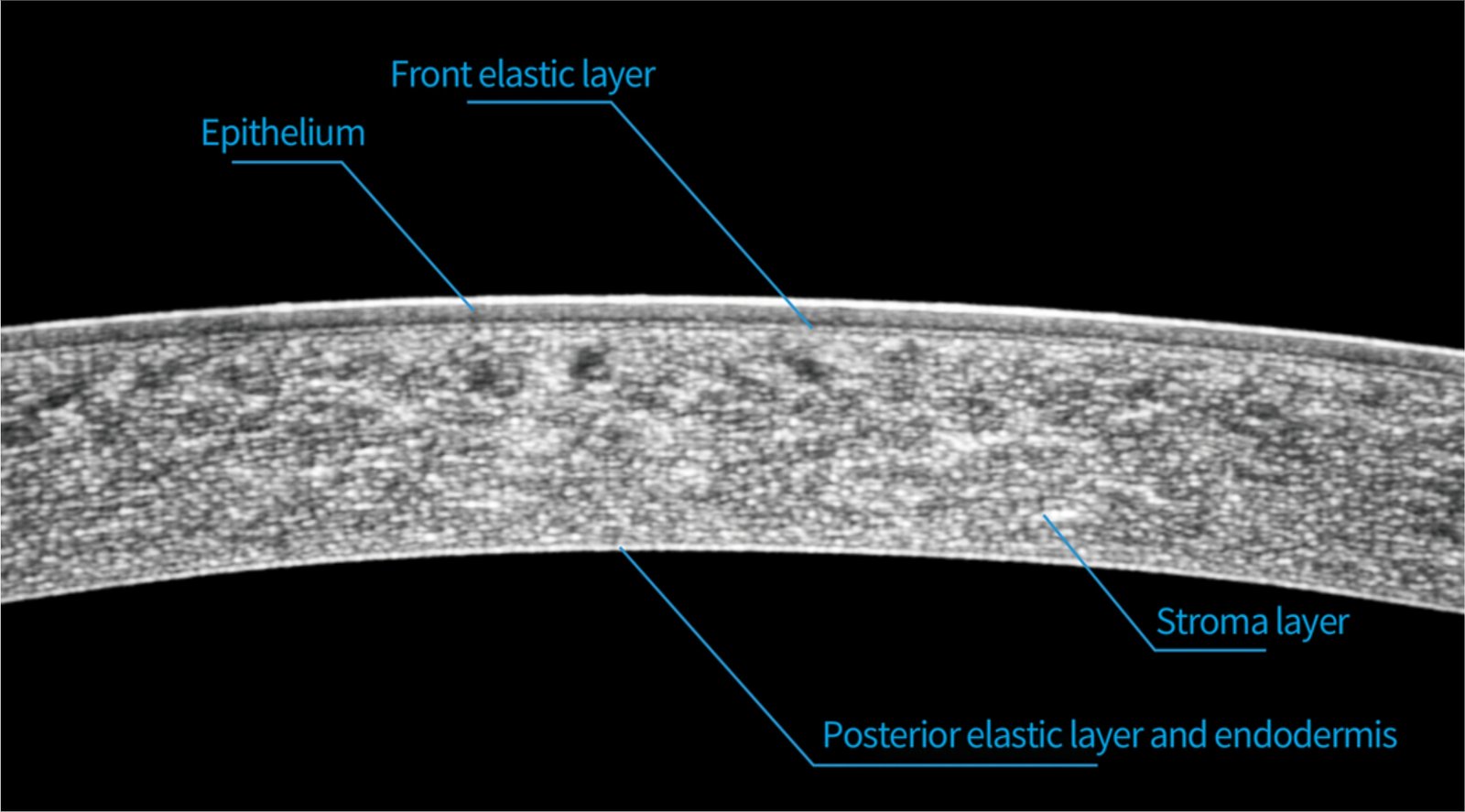
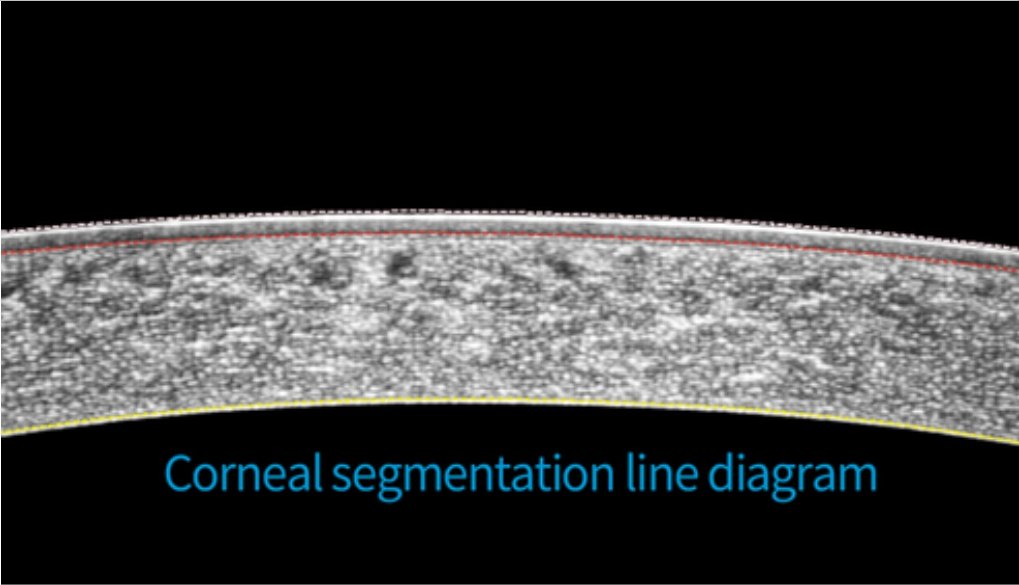
Anterior segment measurement Automatic measurement of corneal thickness, quantification of anterior chamber angle