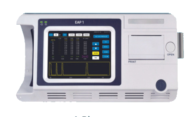
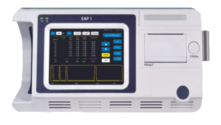
| Specification |
Details |
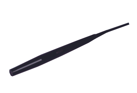
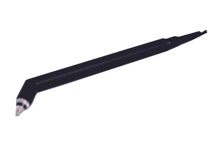
| Probe |
10MHz with Fixation Red Light |
| Total Gain |
100dB with an adjustable range of 0–50dB |
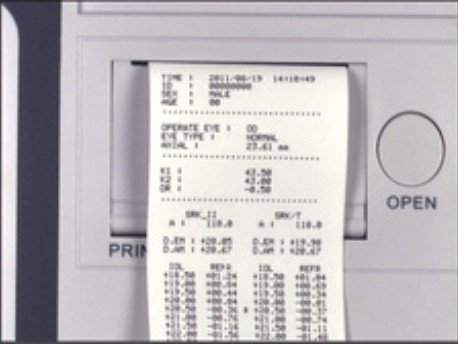
| Biometry Accuracy |
±0.05mm |
| Resolution |
0.01mm |
| Measuring Range |
15–40mm |
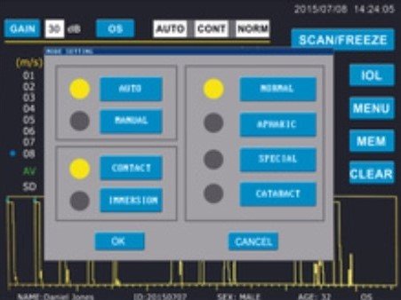
| Measuring Mode |
Contact or Immersion |
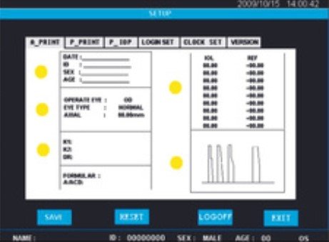
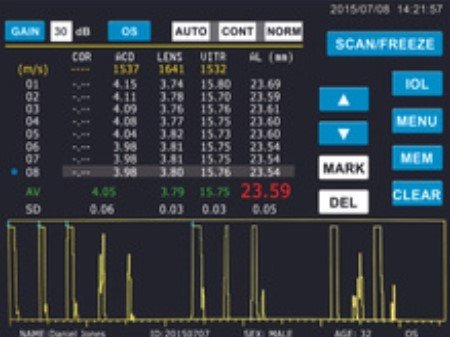
| Measuring Parameters |
Anterior Chamber Depth, Lens Thickness, Vitreous Length, and Axial Length |
| Measuring Modes |
Automatic (Normal, Cataract, Aphakic, and Special) and Manual |
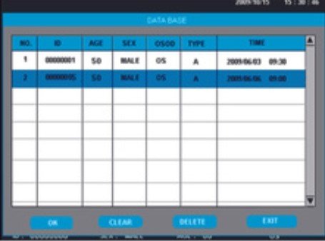
| Groups of Readings |
8 Groups of Readings with Averaging and Standard Deviation |
| Standard Configuration |
| Standard Configuration |
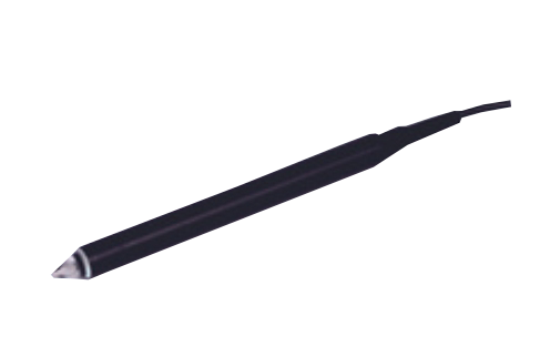
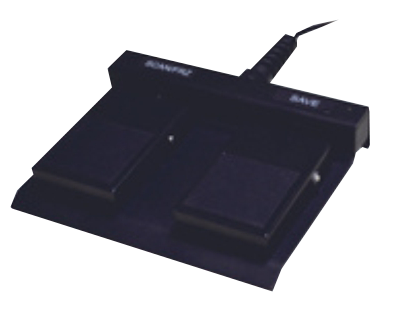
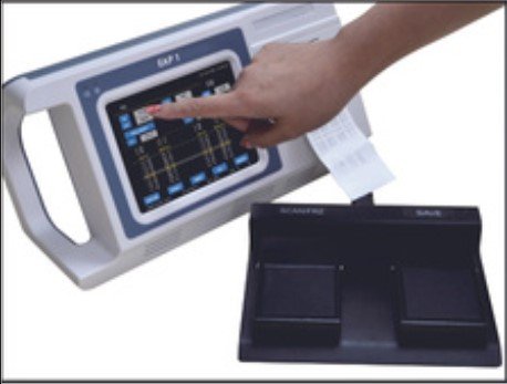
10MHz Probe, 20MHz Probe, Footswitch, Test Object, AC Adapter |
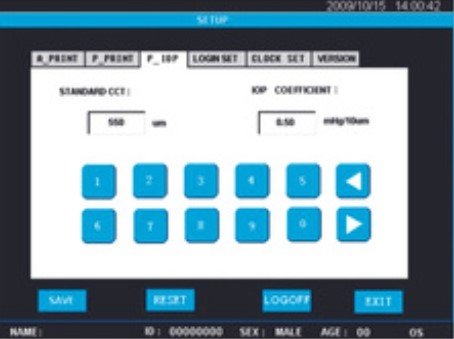
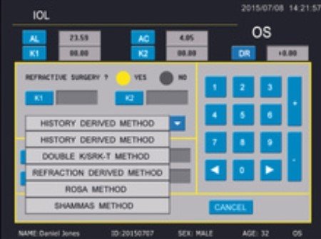
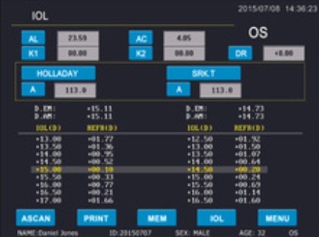
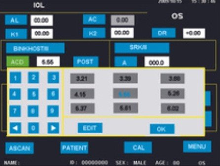
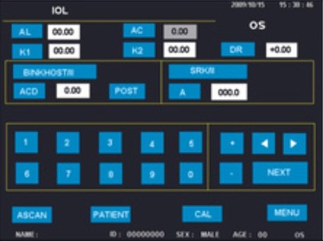
| IOL Calculation |
| General |
SRK-II, BINK-II, HOFFER-Q, History-derived, Refraction-derived, SHAMMAS |
| Post-Refractive |
SRK-T, HOLLADAY, HAIGIS, Double K/SRK-T, ROSA |
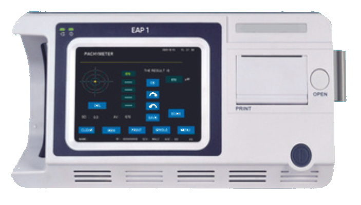
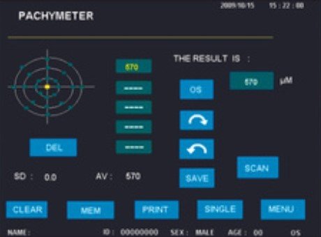
| Pachymeter |
| Probe Frequency |
15–20MHz |
| Display Resolution |
1μm |
| Biometry Accuracy |
±5μm |
| Measuring Scope |
230–1200μm |
| Maps |
Multiple Corneal Maps with Graphical Display |
| General |
| Power Supply |
AC 100–240V, 50/60Hz, 50VA |
| Dimensions |
337mm x 177mm x 155mm (L x W x H) |
| Weight |
1.7Kg |
| Optional |
| Optional |
Immersion Shell |