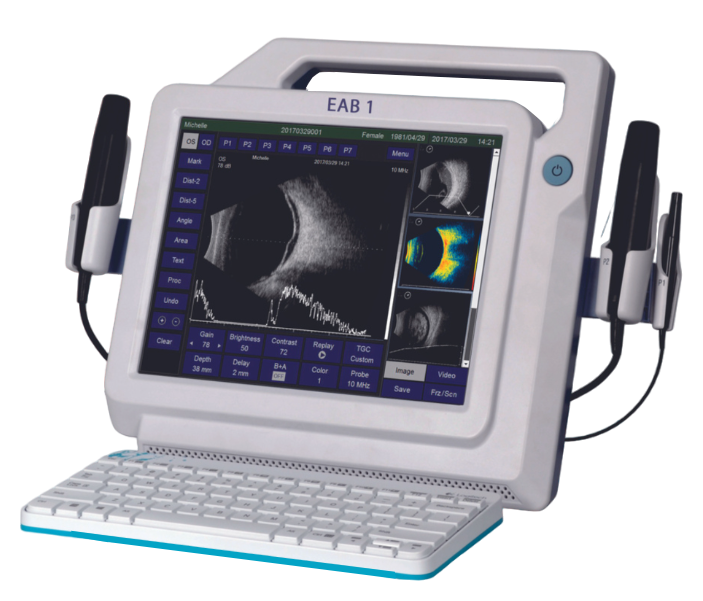
An ultrasonic A/B scanner for ophthalmology is a diagnostic tool used to evaluate the eye’s internal structures through sound waves. It combines A-scan and B-scan ultrasonography
- A-scan (Amplitude Scan): Provides precise measurements of the eye’s length and depth, typically used for determining the axial length for intraocular lens (IOL) calculations, especially in cataract surgery.
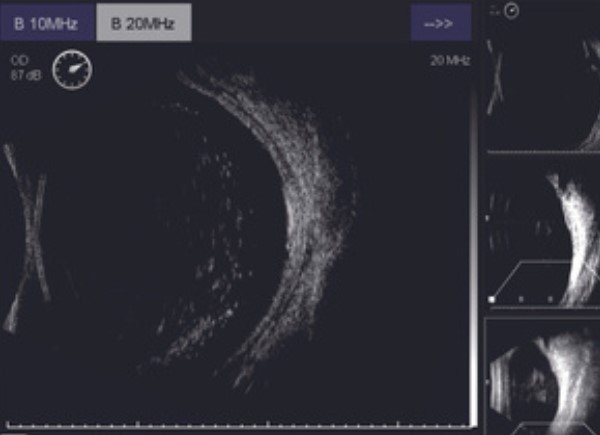
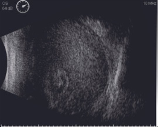
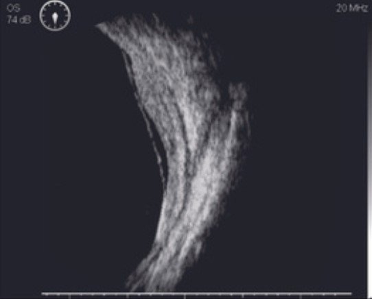
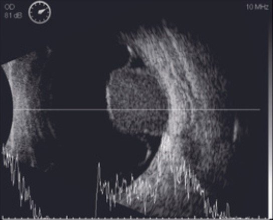
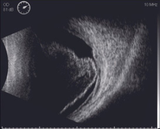
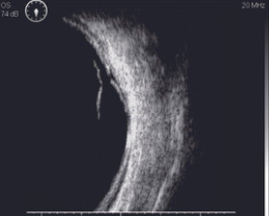
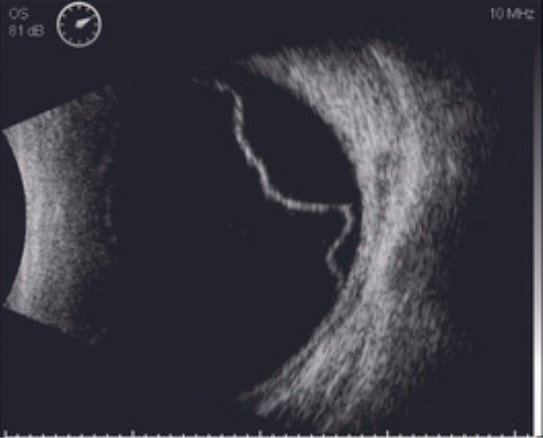
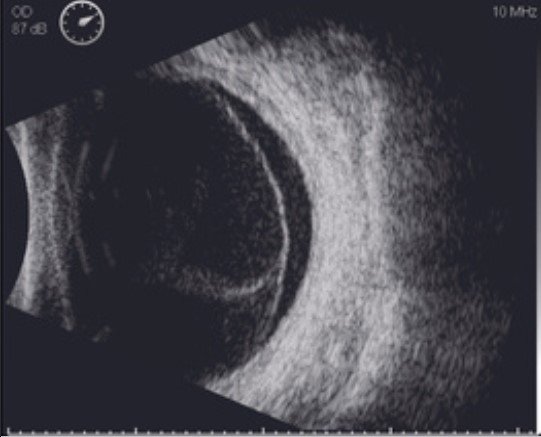
- B-scan (Brightness Scan): Creates a two-dimensional cross-sectional image of the eye and surrounding structures. It’s used to visualize the retina, vitreous, optic nerve, and other parts when direct viewing is obstructed by conditions like cataracts or dense media opacities.
B-Scan Enhanced Visibility and User-friendliness
- Image/video buffer slots for immediate click-and-check, review and comparison
- Images/videos captured in real time and saved in lossless format (.bmp & .avi)
- Zooming and scan depth adjustment for better ocular disease observation
- Vitreous-Enhanced Mode improves visibility at vitreous body

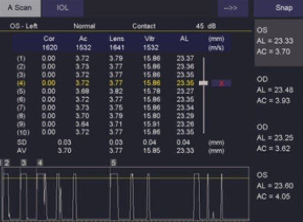
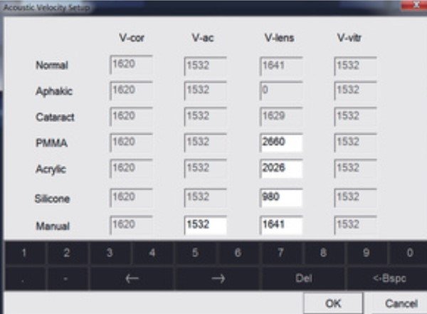
A-Scan & IOL Calculation Enhancements and Tight Integration
- Biometry buffer slots for immediate click-and-check, review and comparison
- Improved accuracy and reliability with Average and Standard Deviation for up to 10 scans per exam
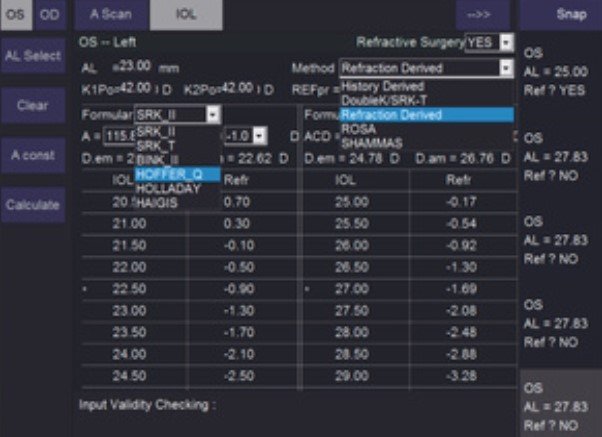
- 6 popular IOL formulae and 5 Post-refractive IOL formulae available
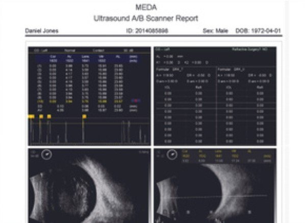
Full Reporting Capabilities
Full report integrating A-scan/IOL results, B-scan images and doctor’s comments with customized dictionary of symptom entries
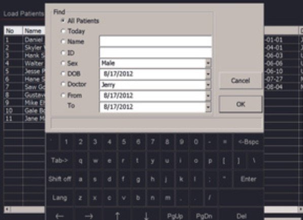
Comprehensive Data Archiving and Management
Simple UI allows users to load, search, print, etc. Unlimited storage capacity for over 20,000 exams (8 lossless images per exam)
Features
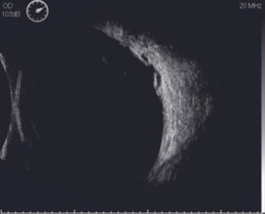
- High Definition 20MHz B-scan Image
- High Resolution LCD touch screen
- Image/video snapped and stored in real time
in multiple buffer slots for immediate comparison and review - Multiple TGC options for operator’s preference including vitreous-enhanced mode
- Editable clinical report with A-scan/IOL results, B-scan images,
and textual comments with configurable entries - Reports in .PDF format for sharing and print-out
- Compatibility with graph/text printer
- Diverse connections via HDMI and USB 2.0 ports
- HDMI interface for double-screen showcase
Specification
| Specification | Details |
|---|---|
| Ultrasound Probes | 10MHz B-Scan Probe, 20MHz B-Scan Probe |
| Axial Resolution | 10MHz B-Scan: <0.1mm, 20MHz B-Scan: <0.08mm |
| Lateral Resolution | 10MHz B-Scan: <0.2mm, 20MHz B-Scan: <0.15mm |
| Scan Depth | 10MHz: 28mm — 60mm, 20MHz: 19mm — 40mm |
| Scan Angle | 53° |
| Cineloop | 10s/100 frames with dynamic replay |
| Image Acquisition | Snapped & saved in real-time without any limitation |
| Gray Scale | 256 Levels |
| Gain | 1-105dB adjustable |
| TGC | Default |
| B+A Mode | Vitreous-Enhanced Mode, Customized |
| Measurements | AL Measurement |
IOL Calculation
| Specification | Details |
|---|---|
| General | SRK-II, SRK-T, BINK-II, HOLLADAY, HOFFER-Q, HAIGIS |
| Post Refractive | History-derived, Double K/SRK-T, Refraction-derived, SHAMMAS, ROSA |
Biometric A-Scan
| Specification | Details |
|---|---|
| Probe | 10MHz with Fixation Red Light |
| Gain | 1-60dB |
| Measuring Method | Contact or Immersion |
| Measuring Range | AL Range: 15mm-40mm |
| Measuring Accuracy | ±0.05mm |
| Measuring Mode | Automatic (Normal, Aphakic, Special, and Cataract) or Manual |
General
| Specification | Details |
|---|---|
| Display | High-Resolution 12.1" LCD |
| Printer Compatibility | Graph/Text Printer and Video Printer (PAL) |
| Interface | Video-Out (PAL), HDMI, USB 2.0 Ports |
| Operations | Touch Screen, Wireless Mouse & Keyboard, Footswitch |
| HDD | 500GB |
| Network | Folder/Report Sharing |
| Power Supply | AC 100~240V, 50/60Hz |
| Optional | 20MHz B-Scan Probe, Eye Cup, Immersion Shell, Video Thermal Printer, DICOM 3.0 Software Package |